
The first global installation of Philips’ Verida Spectral CT at a Madrid university hospital signals a major step forward for spectral imaging and AI-powered diagnostics.
In a hurry? Here are the key takeaways:
- Spain hosts the world’s first installation of the Verida Spectral CTDeployed at Nuestra Señora del Rosario University Hospital in Madrid, it marks a milestone in spectral CT adoption.
- The system integrates spectral imaging and AI across the entire imaging chain.It enables automatic spectral data capture in every scan while improving contrast, reducing noise, and maintaining low radiation doses.
- The installation positions the hospital as a global reference center for advanced imaging.It supports Philips’ broader strategy to expand spectral CT adoption and integrate AI-driven diagnostics into routine clinical workflows.
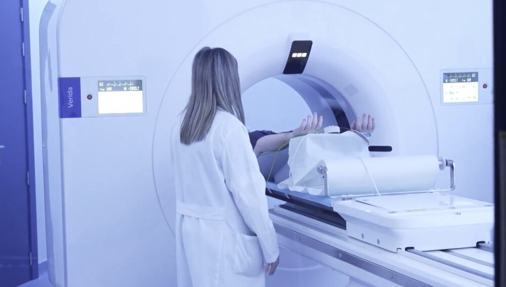
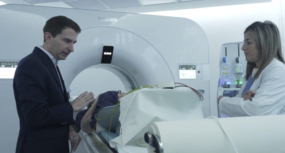
The MRI and CT service at Nuestra Señora del Rosario University Hospital in Madrid has become the first clinical center worldwide to install the Verida Spectral CT system developed by Philips. Announced in March 2026, the installation marks a milestone in diagnostic imaging. It combines detector-based spectral technology with artificial intelligence to deliver anatomical and functional information from a single low-dose scan. The deployment positions Spain at the forefront of clinical imaging innovation and reflects the hospital’s commitment to integrating advanced imaging technologies into everyday clinical practice. For radiologists and hospital administrators alike, the system promises improvements in diagnostic accuracy, workflow efficiency, and sustainability. These areas are increasingly central to modern healthcare delivery.
Verida Spectral CT: Accessible in Routine Clinical Workflows
The Verida Spectral CT represents a new generation of computed tomography designed to make spectral imaging accessible in routine clinical workflows. Spectral CT differs from conventional CT by capturing information across different energy levels of X-rays. It enables clinicians to analyze both anatomical structures and tissue composition.
In many traditional spectral systems, specialized protocols or additional post-processing steps are required. Verida was designed to remove that complexity. Its spectral capabilities operate automatically across all scans. It allows clinicians to obtain spectral information without altering their standard workflow.
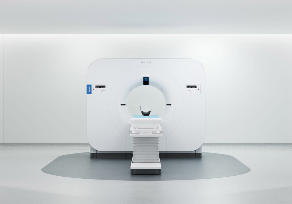
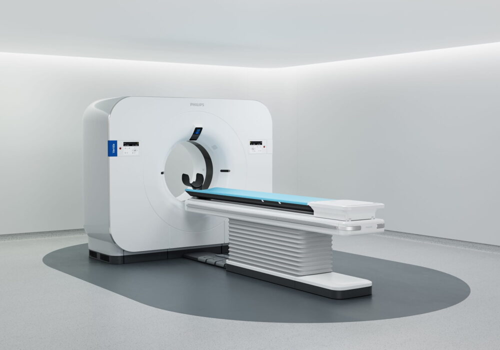
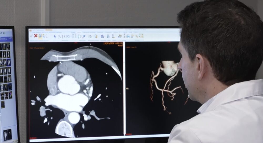
At the core of the system is Philips’ third-generation NanoPanel Prism Precise detector paired with the Spectral Precise Image reconstruction engine. Together, these technologies integrate artificial intelligence across the imaging chain, from acquisition to reconstruction, producing spectral datasets that radiologists can analyze immediately.
According to Philips data, AI-powered spectral reconstruction can deliver up to 136 percent more contrast while reducing image noise by as much as 80 percent, all while maintaining low radiation dose levels. Such improvements may enhance the detection of subtle lesions and improve tissue characterization, which are critical factors in oncology, cardiology, neurology, and emergency imaging.
Another key advantage lies in the system’s “always-on” spectral capability. Because spectral data are captured during every scan, clinicians can retrospectively analyze tissue composition without repeating the examination. This may reduce the need for follow-up imaging or additional diagnostic procedures, potentially accelerating clinical decision-making and improving patient pathways.
Performance was also designed with high-demand radiology departments in mind. The system supports reconstruction speeds of up to 145 images per second and can handle up to 270 studies per day in high-volume configurations. Such throughput is increasingly important as healthcare systems face rising patient demand and limited imaging resources.
Importantly, Verida integrates directly with hospital imaging infrastructures through PACS systems using Philips’ Spectral Magic Glass interface. This allows radiologists to access spectral information within their existing viewing environment rather than requiring separate workstations.
Integration at the Madrid Hospital for Patient Safety
For the MRI and CT service at Nuestra Señora del Rosario University Hospital, the installation of Verida represents both a technological milestone and a strategic step toward expanding advanced diagnostic capabilities.
The hospital’s radiology department has long emphasized innovation in imaging technology, and the integration of spectral CT aligns with that objective. By introducing spectral imaging into routine workflows, the center aims to enhance diagnostic confidence while maintaining operational efficiency.
Dr. Eliseo Vañó, head of the radiology service at the hospital, emphasized the clinical value of the system in everyday practice.
“The Verida Spectral CT allows us to naturally integrate spectral imaging into our daily work without disrupting the reading workflow or adding operational burden,” he said. “Having functional and anatomical information in a single study, with high image quality and low dose, improves patient safety and strengthens our ability to reach accurate diagnoses from the outset.”
For clinicians, the ability to access spectral information automatically may improve the assessment of complex cases. Tissue characterization, lesion detection, and vascular evaluation are among the areas expected to benefit from enhanced spectral analysis.
The system may also contribute to more efficient patient management. By reducing the need for repeat imaging or additional tests, spectral CT can help streamline diagnostic pathways while minimizing patient exposure to radiation.
Operational sustainability was another factor in the system’s design. Verida incorporates AI-based remote services that enable proactive monitoring of system performance and optimization of energy consumption. According to Philips, this architecture can reduce energy use by up to 45 percent while improving X-ray tube management and extending system lifespan.
For hospital administrators, such efficiencies can translate into lower operational costs and more sustainable management of healthcare resources. This is an increasingly important consideration as hospitals seek to balance innovation with financial and environmental responsibility.
The Next Steps for Philips and Spectral CT
The installation of the Verida Spectral CT in Madrid represents an early step in a broader strategy by Philips to expand the adoption of spectral CT technology worldwide.
Spectral imaging has long been recognized for its potential to improve diagnostic precision, but adoption has historically been limited by workflow complexity and technical barriers. By embedding spectral functionality directly into the imaging chain and automating the process with AI, Philips aims to bring spectral CT into routine clinical practice.
The deployment in Madrid is likely to serve as an early reference site for the technology. As clinical teams gain experience with the system, their findings may contribute to further research and validation of spectral CT’s clinical benefits.
At the same time, the broader imaging industry is moving toward more intelligent and integrated diagnostic platforms. Advances in detector technology, artificial intelligence, and imaging reconstruction are enabling systems that not only produce higher-quality images but also provide deeper insights into tissue composition and disease processes.
For radiology departments worldwide, these developments point toward a future in which advanced imaging technologies—once reserved for specialized centers—become standard tools in everyday clinical practice.
The first installation of Verida at Nuestra Señora del Rosario University Hospital, therefore, represents more than a single technological milestone. It reflects the evolving role of radiology as a central driver of precision medicine and data-driven healthcare.