
New PET and SPECT radiotracers targeting amyloid proteins could enable earlier, non-invasive diagnosis of cardiac amyloidosis and support the shift toward precision diagnostics.
In a hurry? Here are the key takeaways
- Bayer is entering molecular imaging.With the acquisition of investigational PET and SPECT amyloid tracers designed to diagnose cardiac amyloidosis earlier and potentially reduce the need for biopsy.
- The lead PET tracer (ATO1) is nearing the end of Phase III development.It has received FDA Breakthrough Therapy Designation, signaling potential for accelerated regulatory review.
- Molecular imaging will complement—not replace—MRI and CT.It will provide biological insight into disease processes and support more precise diagnostics and treatment decisions.
Following its acquisition of Attralus, Bayer is expanding its diagnostic imaging portfolio into molecular imaging with two investigational amyloid-targeting radiotracers. The compounds, including a PET tracer currently in late-stage Phase III development, are designed to detect amyloid protein deposits associated with cardiac amyloidosis and other systemic diseases.
The move marks a strategic step beyond Bayer’s traditional expertise in contrast-enhanced CT and MRI imaging. While anatomical imaging technologies remain the backbone of clinical diagnostics, molecular imaging enables physicians to visualize biological processes underlying disease. According to Bayer researchers, integrating these two approaches could improve diagnostic certainty and help guide more targeted therapies for patients.
Molecular Imaging Agents Aim to Improve Diagnosis of Cardiac Amyloidosis
The compounds acquired by Bayer target amyloid deposits that accumulate in tissues in diseases collectively known as amyloidosis. One of the most serious forms, cardiac amyloidosis, occurs when abnormal protein fibrils infiltrate the heart muscle, eventually leading to heart failure.
Currently, diagnosing amyloidosis can be challenging. Imaging techniques such as echocardiography or MRI can reveal structural changes in the heart that suggest the disease, but they cannot definitively confirm the presence of amyloid deposits. In many cases, physicians still rely on tissue biopsy for confirmation.
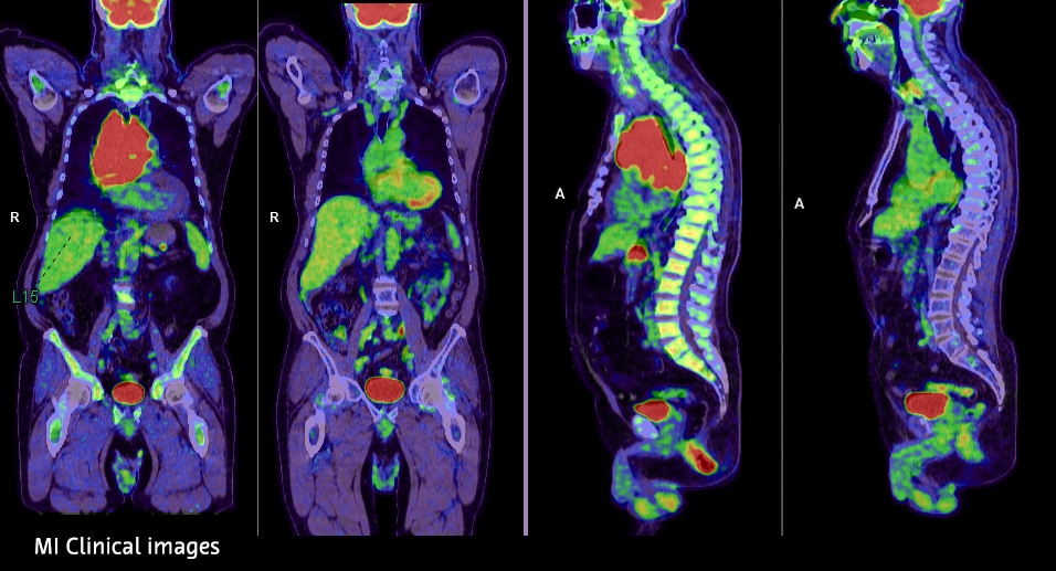
The newly acquired molecular imaging agents are designed to address this gap. The leading compound, known as ATO1, is a positron emission tomography (PET) tracer that uses the radioisotope iodine-124 to bind to amyloid proteins. When injected into the patient, the tracer accumulates in areas where amyloid is present, allowing physicians to visualize deposits during a PET scan.
“What we know is that standalone MRI or echocardiography can show signs that are indicative of cardiac amyloidosis, but they can’t definitively say that amyloidosis is there. The only way to do that today is with a biopsy,” Dr. Konstanze Diefenbach, Head of R&D Radiology at Bayer, explained in an interview with MedicalExpo e-Magazine during ECR 2026. “To have a diagnostic agent that definitively says yes or no adds an additional layer of certainty on top of anatomical imaging.”
The tracer is designed as a pan-amyloid agent, meaning it can detect multiple subtypes of amyloidosis rather than targeting only a single form of the disease. This could be particularly important because current nuclear imaging tests are effective for only certain amyloid subtypes, leaving many patients without reliable non-invasive diagnostic options.
Alongside the PET tracer, Bayer is also developing a second compound, ATO5, designed for single-photon emission computed tomography (SPECT). While SPECT imaging provides less quantitative detail than PET, it is widely available and generally less expensive. Researchers believe the two agents could ultimately serve complementary roles in clinical practice, balancing diagnostic precision with broader accessibility.
Integrating Molecular Imaging Into Clinical Practice
Despite the introduction of new tracers, Bayer researchers emphasize that molecular imaging will not replace existing diagnostic imaging modalities. Instead, it will complement them by providing additional biological insights that anatomical imaging alone cannot deliver.
“We’re adding something on top,” Dr. Konstanze Diefenbach said. “CT and MRI remain the workhorses of medical imaging. They help us understand anatomy and morphology. But in certain areas, you want to understand the biology behind what you’re seeing.”
Modern imaging systems already combine these approaches. PET scanners are typically integrated with CT or MRI, allowing clinicians to capture anatomical and molecular information in a single imaging session. During the scan, patients remain on the same scanner table while the two datasets are acquired sequentially and combined.
Researchers say this integrated approach could significantly simplify the diagnostic journey for patients suspected of having amyloidosis. Today, patients often undergo multiple tests—including echocardiography, MRI, nuclear imaging, and sometimes biopsy—before receiving a definitive diagnosis.
Amyloidosis symptoms are often nonspecific and can resemble other forms of heart disease. As a result, diagnosis frequently occurs late in the disease course, when patients have already developed severe heart failure.
“With such a tracer, once it is established in the general guidelines, clinicians could go directly to this tool when they suspect amyloidosis,” Dr. Erin Bowman, Medical Affairs Lead for Molecular Imaging at Bayer, said. “That could help simplify the pathway and detect patients earlier.”
Earlier diagnosis could also carry significant economic and clinical benefits. Amyloidosis therapies can be costly, and patients often experience repeated hospitalizations as their disease progresses. Identifying the condition sooner could allow physicians to initiate treatment earlier and potentially reduce long-term healthcare costs.
“The goal is not to add burden to the patient,” Dr. Erin Bowman noted. “It’s to provide a tool that helps the patient and the healthcare provider optimize care.”
Phase III Development and the Path Toward Global Approval
The ATO1 PET tracer is currently nearing the end of Phase III clinical development, the final stage of testing before regulatory submission. Phase III studies typically involve large patient populations and are designed to confirm both safety and diagnostic performance. Researchers say early results appear promising, though final data analysis is still ongoing.
“So far, the signals look very good, but that’s exactly why we conduct clinical trials,” Dr. Erin Bowman said. “You need the full dataset before moving forward with regulatory discussions.”
The compound has already received Breakthrough Therapy Designation from the U.S. Food and Drug Administration, a regulatory pathway intended to accelerate development of technologies that may offer substantial improvements over existing options.
Beyond clinical validation, Bayer is also working to establish the manufacturing and supply chain infrastructure required for radiopharmaceutical production. Because imaging tracers contain radioactive isotopes with relatively short half-lives, reliable manufacturing and distribution networks are essential.
“This is not an easy task,” Dr. Konstanze Diefenbach acknowledged. “You need consistent production, quality standards, and a reliable supply chain to bring the compound to hospitals.”
Once these elements are in place, Bayer plans to pursue regulatory approvals in major markets including the United States, Europe, and Asia.
While the exact timeline remains uncertain, researchers suggest the program is already approaching the stage where regulatory conversations can begin.
“We are not ten years away,” Dr. Erin Bowman said.“We are at the point where we are preparing the data, preparing manufacturing, and thinking about how to approach regulatory authorities.”