
Dr. Robert Kerstein, DMD, Computerized Bite Analysis Expert at RBR Consulting, explains how T-Scan is replacing paper and ink subjective bite adjusting for patient bite care.
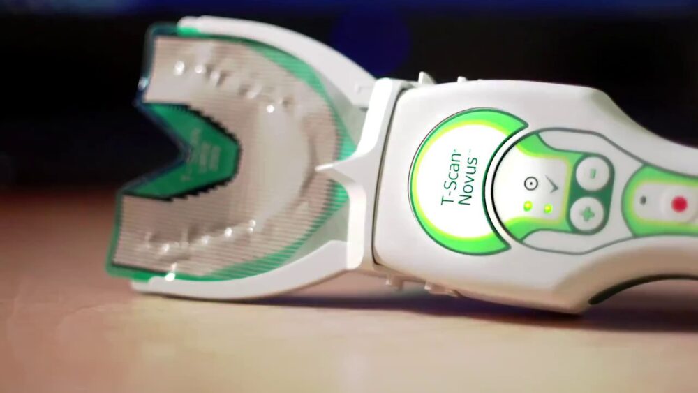
The T-Scan™ 10 technology from Tekscan Inc., has revolutionized dental bite procedures by objectively measuring patient bite forces and timing, which drastically improves bite problem diagnosis and treatment. T-Scan data can guide a dentist to make accurate, high-precision bite adjustments that optimize the lifespan and comfort of dental restorations while also improving patient well-being.
T-Scan recordings detect which forceful tooth contacts need bite force controlling adjustments, and which healthy low force contacts should be left alone. Compared to the outdated articulating paper and ink subjective method, T-Scan is minimally invasive, and protects patients from dentists guessing poorly at paper and ink marks that do not measure any bite function.
The coming years will determine whether T-Scan transitions from a minimally adopted, long-standing available dental innovation to a widely recognized therapeutic modality. Dentistry will only advance the quality and safety of patient bite care by replacing the paper mark subjective method with T-Scan’s force and timing data to accurately and safely adjust patient bites.
The Connection Between Bite Adjustments and Common Health Conditions
Multiple published studies and Digital Occlusion textbook chapters have shown T-Scan-guided bite adjustments can improve common health concerns and symptoms including:
- Decrease TMJ muscle pain and tension
- Strengthen chewing
- Reduce headaches
- Reduce bruxing/clenching habits
- Reduce medication need/use
- Reduce unnecessary bite adjustment visits following dental procedures
- Statistically lessen Implant complications like chipping porcelain and fracturing implant parts
- Improve posture
- Increase airway size
- Reduce stress and cortisol levels to improve emotional well-being.
These patient improvements occur without the need for splints, nightguards, orthotics, CPAP, Botox injections, TENS unit, medications, or physical therapy.T-Scan-guided bite treatment is so precise, it therapeutically works from within the patient’s central nervous system, to control how teeth neurologically interact with the center of the brain. T-Scan exerts direct neural control the over the volume of electrical output coming from one’s back teeth, eliminating the need for external symptomatic treatments for most patients.
Unfortunately, many common health conditions are often medically misdiagnosed or managed in isolation because they present without clear signs of organic disease (no tumors, cysts, or infections) and are rarely considered “bite-related.”
As a result, patients may spend years treating symptoms without anyone evaluating whether occlusion is contributing to the problem. The following conditions and symptoms are among those that can, in some cases, be influenced by bite dysfunction:
- Temporomandibular Disorders Symptoms (TMD)- Facial Pain and tension, chewing fatigue, frequent jaw soreness, reduced mouth opening, clicking, popping and locking TM joints, unilateral facial swelling, clenching and Bruxism
- Some Hearing Disorders – Tinnitus, Meniere’s Disease (progressive unilateral hearing loss), unexplained recurrent earache, ear fullness, vertigo, drop attacks
- Trigeminal Neuralgia – Episodic shooting or burning facial pain
- Chronic tension headaches – and some migraine headaches
- Posture Anomalies – Forward Head Posture (FHP), uneven shoulder alignment, head canting
- Restricted Neck Motion –turning limitations, shoulder and arm pain, throat tightness
- Brain Fog – Episodic cloudy consciousness
- Airway constriction – A component of Obstructive Sleep Apnea (OSA)
- Some Ticks and Dystonias – Unexplained eye twitching, and intermittent facial, neck, and shoulder ticks are often bite-related
Too often, these conditions are ineffectively addressed using symptomatic management alone. Interventions such as mouthguards designed to separate teeth or open the airway, physical therapy, laser therapy, acupuncture, repeated pain medication use, or frequent Botox injections may provide temporary relief by reducing muscle tension or discomfort. However, symptoms often return because these approaches do not target the underlying bite imbalances.
Emerging research and clinical experience show that T-Scan–guided, high-precision bite adjustments can help reduce many of these complex symptoms by improving occlusal stability and influencing neuromuscular function. By targeting the root mechanical imbalance rather than solely managing outward symptoms, clinicians may achieve more durable and comprehensive patient outcomes.
How Traditional Articulating Paper Puts Patients at Risk for Future Bite Problems
What’s unfortunate for patient safety and bite care quality is that the subjective paper mark method is still used worldwide as a reliable and safe method despite studies citing its inaccuracy. This continued use of an outdated method remains a top driving force as to why bite complications arise after new dentistry is installed.
What patients don’t realize is that a dentist without T-Scan will often adjust the wrong places on their teeth or implants by using the outdated paper mark method. It’s based on “ink mark sizes” and not on true bite force levels. Paper mark “size” only correlates to bite force levels 14% of the time. That means, if a dentist chooses a “big ink mark” to adjust thinking, it’s a high force bite contact, it will only be high force 14% of the time. Conversely, if the same dentist thinks “a little mark is low force”, it will only be low force 14% of the time. This means a dentist choosing contacts by size will adjust the wrong contacts 86% of the time. This inaccuracy creates underlying bite problems after new fillings are placed, a crown or two is placed, or even after an Invisalign treatment. The imbalanced bite results in headaches, jaw or tooth pain, or a host of other symptoms that unexpectedly emerge.
This all-too-common problem is rampant and uncontrolled in dentistry and has not been addressed by the ADA or any dental school. The paper mark method is still being taught worldwide, as if the size of the ink marks MEASURES bite forces, when multiple studies clearly show ink size and paper marks are incapable of measuring anything about the bite, except where top and bottom teeth touch.
Patients Should Make Sure to be Treated by a Properly Trained T-Scan Clinician
Despite dentistry advancing with the ability to fabricate crowns and implants, the digital workflow can’t control the bite forces. It is essential for a patient to bite and grind on a T-Scan sensor with their new veneers or implanted teeth to fully capture the bite force problems and guide any needed force-controlling adjustments. This is the only way to accurately and predictably alter the new bite on new dentistry.
Much like any technology, patients should understand that just because a dentist“owns a T-Scan”, it doesn’t mean that the dentist is an effective and knowledgeable T-Scan user. Dentists need definitive clinical T-Scan training with patients overseen by a knowledgeable T-Scan mentor, educating them to correctly record patient bite function, how to understand that data, and how to implement that data to guide adjustments.
When identifying a dental provider, patients should seek out those who utilize the technology while also inquiring on their training prior to scheduling an appointment. To find a list of properly trained and educated T-Scan clinicians go to: www.digitalocclusionseminars.com/dtr-providers.html

Dr. Robert Kerstein, DMD, Computerized Bite Analysis Expert at RBR Consulting
Dr. Robert B. Kerstein received his D.M.D. degree in 1983, and his Prosthodontic certificate in 1985, both from Tufts University School of Dental Medicine. From 1985 – 1998, he maintained an active appointment at Tufts as a clinical professor teaching Fixed and Removable Prosthodontics. In 1984, Dr. Kerstein began studying the original T Scan I technology, and has since that time has also studied T-Scan II, T-Scan III, T-Scan VII, T-Scan 8, T-Scan 9, and now the present-day version, the T-Scan 10 Novus technology.