A new partnership between Case Western Reserve University in Cleveland (USA) and Microsoft, and centered on the software giant’s HoloLens mixed reality head-mounted display could significantly change how anatomy is taught.
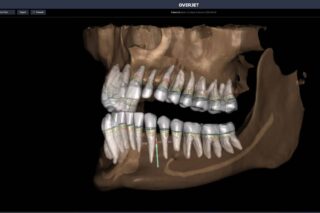
This holographic computer is worn like glasses. It allows professors and students to see in front of them the entire human body in 3D, to move it around and to remove and replace parts. This system of mixed reality—virtual reality superimposed on the real world—offers a view of the nervous system, the skeletal structure, the different organs and even blood circulation. There is no screen to touch or mouse to click. The user simply gestures to create, shape and size the holograms.
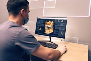
According to Microsoft, HoloLens makes it possible to “bring a digital world into a real world.” Built on Windows 10, its combination of specialized components enable holographic computing. The optical system works in coordination with sensors, and the device contains more computing power than the average laptop. It is also completely untethered—no wires, external cameras, phones or PC connections are needed. Students can use it in class to clearly see muscles overlying the skeleton. They can render the body translucent to look inside and understand cardiac anatomy.
A Case Western student who took part in the HoloLens project explains on the university website that when he saw the aortic valve in 3D in front of his eyes, he understood for the first time where it was and how it really worked. According to Mark Griswold, Case Western Professor of Radiology who experimented with the prototype, this technology could “improve students’ confidence in learning anatomy” without the stress of learning on a cadaver.