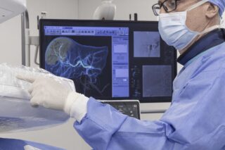
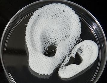
The Wake Forest Institute for Regenerative Medicine reported in mid-February in Nature Biology successfully printing tissue with a custom-designed 3D printer, called ITOP (Integrated Tissue and Organ Printing System). The structures proved to be functional when implanted in animals. MedicalExpo talked to Dr. Anthony Atala, director of the institute and senior author of the study.
MedicalExpo: Dr. Atala, you have printed tissue before. How is your latest accomplishment a breakthrough?
Dr. Anthony Atala: We started research on 3D printing human-scale tissues twelve years ago. Finally, we can print structures that can be implanted surgically, that survive outside a laboratory and that have the structural integrity to function once implanted. We have a solution to the challenge of providing the printed tissue with the nutrition it needs and therewith to ensure that the implanted structures live long enough to integrate with the body.
MedicalExpo: How did you make it functional?
Dr. Anthony Atala: We created microchannels that go into the structures. These microchannels are like highways that allow the nutrients and oxygen from the body to get to the central portion of the structure, so that it gets the nutrition it needs. The implants can then develop a system of blood vessels.
MedicalExpo: Have you tried it in humans?
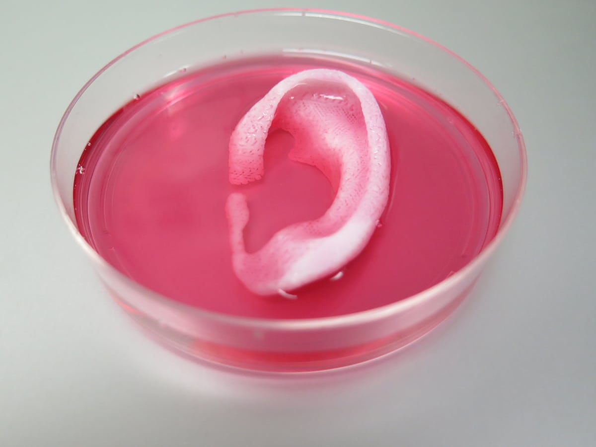
Dr. Anthony Atala: No. We developed a wide range of tissues in terms of their strength. We printed soft tissue, which is muscles. We printed medium-strength tissue, which is cartilage. And we printed strong structures, which are bones. This was to show that the printer is able to print all of them. We implanted them in mice and rats. Months later, blood vessels had formed.
MedicalExpo: When will you be able to implant printed tissue in humans?
Dr. Anthony Atala: We aim to implant bioprinted muscle, cartilage and bone in patients in the future. Right now, we are working on the safety studies to [enable us to plan] for the future. You know that in science it is hard to predict.
MedicalExpo: Can you describe the process from planning to printing the final part?
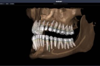
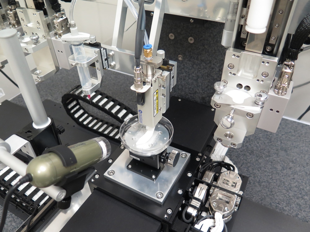
Dr. Anthony Atala: We start with an inventory of the defect area. We then download the digital information from the x-ray into our software program that drives the printer. Finally, it prints the structure that fits into the patient.
One advantage of our ITOP system is that we can use data from CT and MRI scans to plan customized tissue for patients. The actual tissue is made of biodegradable, plastic-like materials to form the structure and of water-based gels with the cells. Those come from the same patient so that there is no rejection.
MedicalExpo: What are the implications of your results for tissue donation?
Dr. Anthony Atala: Growing replacement tissues and organs in the laboratory should help to overcome the shortage of donated tissue for transplants. Because we produce from the patient’s own cells, the goal is to reduce the tissue waitlist for patients.