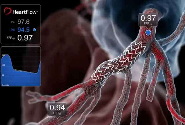
Patients with life-threatening coronary heart disease will be diagnosed and treated five times faster thanks to a new revolutionary tool that uses deep learning technology. It turns a regular CT scan of the heart into a 3D image allowing doctors to diagnose heart disease in just twenty minutes, reducing reliance on unnecessary invasive diagnostic procedures such as angiograms.
The technology, HeartFlow, is already used in 50 heart hospitals in the US as well as across Europe and Japan. It is now being rolled out in the UK to treat around 100,000 patients suspected to have heart disease. The tool takes data from a coronary CT angiography (CTA) scan and uses deep learning technology as well as highly trained analysts to create a personalized, digital 3D model of the patient’s coronary arteries. The algorithms solve millions of equations to simulate blood flow in a patient’s arteries to help clinicians assess the functional impact of any blockages.
Once a patient is diagnosed using the 3D image, treatments include surgery, medication or having a stent fitted. For less serious cases, patients will be given tips on healthy lifestyle changes or cholesterol-lowering medication—meaning the risk is quickly resolved before it becomes life-threatening. The technology could help tackle the overwhelming backlog of patients that has built up due to the disruption to health services across the globe during the Covid-19 pandemic.

Some 17.9 million people die each year from cardiovascular disease—an estimated 31% of all deaths worldwide. The most common type is coronary heart disease (CHD). Known as angina, CHD occurs when the coronary arteries become narrowed by a build-up of fatty material within their walls reducing the supply of oxygen-rich blood to the heart muscle. It develops slowly over time and the symptoms can be different for everyone. They include chest pain, shortness of breath, pain traveling through the body, feeling faint and nausea.
Avoiding Unnecessary Invasive Procedures
There are several tests used to diagnose angina. An electrocardiogram (ECG) detects problems with heart rate or rhythm and is an indicator of whether a heart attack has occurred. It is usually one of the first heart tests carried out but has limitations so further testing is normally needed. An angiogram is a special type of X-ray which uses contrast dye to assess the blood flowing to the heart. The procedure involves local anesthetic to insert a catheter into the groin, which is then directed up to the heart. After it is done the patient will be put on bed rest for an hour or so and in some cases overnight if the wound is painful.
HeartFlow technology means that patients can be assessed swiftly and painlessly without needing bed rest. Around 100,000 people in the UK with recent onset chest pain who are offered coronary CT angiography will now be eligible for HeartFlow over the next three years, with more than 35,000 people set to benefit each year. Professor Sir Nilesh Samani, Medical Director at the British Heart Foundation, said:
“This technology will prevent unnecessary admissions for angiograms and quickly provide information that allows patients to be put on the best treatment pathway for their condition.”
Dr. Derek Connolly, a consultant interventional cardiologist at Sandwell & West Birmingham Hospitals NHS Trust, added:
“For every five patients who have a cardiac CT and a HeartFlow Analysis, four patients go home knowing they don’t need anything else. Half of those patients will be on cholesterol tablets because they have early disease, and the other half will have normal coronary arteries. Incorporating the HeartFlow Analysis has had a meaningful impact at our hospitals, improving the diagnosis and treatment of the leading cause of death.”


