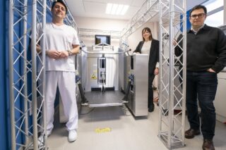
A team from Carnegie Mellon University in the US has succeeded in printing a 3D model of a human heart which looks and feels like real cardiac tissue, using a material derived from seaweed.
In the short term, the Freeform Reversible Embedding of Suspended Hydrogels (FRESH) technique aims to create replica organs to enable cardiac surgeons to visualize and plan complicated procedures with more precision than ever before. Adam W. Feinberg, Professor of Biomedical Engineering and Materials Science and Engineering at the university, said:
“One day, the hope is that it might be suitable for creating replica body parts for transplant. Our long term goal is to be able to FRESH 3D bioprint a heart for transplant, but that is decades off.”
He added:
“However, we hope to be able to FRESH print parts of the heart, such as valves and sections of the ventricle, much sooner and have a major impact in that way in a matter of years.”
Life-Like Tissue
The team has been working on 3D bioprinting for nearly 10 years now, with the aim of being able to print tissue and organs to repair the human body following injury or disease. The FRESH printing technology was developed to solve the problem of 3D printing soft materials and cells. Professor Feinberg said:
“Over the past five years, hospitals have greatly expanded their use of 3D printed models of organs for patient education, surgical training and surgical planning. We realized about two years ago that our technology could make a much more immediate impact by creating life-like tissue and organ models that would improve the realism over these rigid plastic models.”

In 2018, the team started working on printing a life-size heart. He explained:
“This required building new 3D bioprinters, because this is probably the largest bioprinted hydrogel scaffold ever created, and it required a bigger build volume and a lot of bioink. The alginate we use is a hydrogel that is widely used in the biomaterials and tissue engineering fields. It is a great material for creating models because it is relatively inexpensive for a bioink but has tissue-like mechanical properties.”
Beyond the Heart
All surgical models have the advantage of helping visualize and plan the surgery. They also have the added benefit that they can be cut and sutured like real tissue. The team can currently make a model of anything they have the 3D file for, including babies’ hearts. Professor Feinberg said:
“Currently we use MRI images of the human heart, and we can also use MRI images from pediatric patients. We are now in the process of working with surgeons on our models and will use that feedback to improve them. The next stage in development is working with surgeons to improve the feel and performance of the model, and to expand to tissues and organs beyond the heart.”


