
With a variety of options available, deciding which microscope camera best fits your experimental needs can be daunting. If you’re trying to decide, make sure you check out this camera selection guide from Leica Microsystems which outlines the key factors to consider.
By Leica Microsystems.
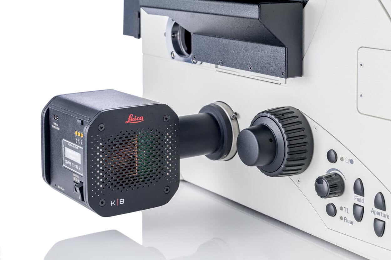
Microscope Cameras for Life Science Research
Cameras are a vital part of microscopy systems, converting images into digital data, and they can have a significant impact on what you can achieve with your system. From basic workflows such as monitoring cell confluency and documenting stained tissue sections, through to challenging high speed live-cell experiments, cameras are required to capture reliable, reproducible, and quantifiable data for downstream analysis to yield publishable data.
Factors to Consider When Selecting a Microscope Camera
Thinking about a few different aspects related to cameras can help you obtain images with the optimum amount of information for your research. Before diving into the technical specifications of different cameras, it’s important to first carefully consider your samples and the techniques you will be performing.
The contrast methods you need will also have an influence on your choice of camera, as will the type of data you’re ultimately hoping to collect from your sample. In this guide, we start by reviewing these considerations before going on to clarify some of the technical aspects.
Technical Aspects
The requirements for microscopy cameras are application specific. Simply having plenty of megapixels will not often deliver the best image. Factors like pixel size, noise levels, dynamic range, and frame rate are also important factors to consider.
It is important to remember that different factors are often interdependent. For example, sensitive cameras often have larger pixels, which can mean lower resolution, which could be suboptimal for some applications.
Conversely, large pixels typically offer greater dynamic range which can be highly advantageous for brightfield imaging. It is therefore very important to consider what is most important to you and to choose an optimal solution for your application. We discuss these points further in this guide and then look at what makes a camera good for brightfield vs. fluorescence imaging.
Download the Camera Guide to Find Out:
- What makes a camera good for fluorescence imaging or brightfield imaging, or both!
- What the technical considerations for camera selection are, with terms such as frame rate, quantum efficiency, bit depth, dynamic range, and much more, explained.
- How to decide which camera is right for your life science microscopy experiments.
- Which Leica Microsystems camera may be best for you.
Why Choose a Microscope Camera From Leica Microsystems?
Leica offers a range of microscope cameras which use the latest generation of modern CMOS sensors to address a wide variety of application needs—even demanding fluorescence applications. Since a camera is only part of the overall microscope, its integration into the system will ultimately determine what you are able to achieve with it.
Your Benefits With Leica Solutions Include:
- Software: Leica’s LAS X and Enersight software packages seamlessly integrate their cameras into the microscope system.
- Control: Operate your camera in multiple readout modes, enabling the configuration that best matches your application needs.
- Flexibility: Features such as image and hardware-based auto-focus to keep your specimen in view, combined with auto exposure and flexible experimental design tools, enable you to perform a wide variety of tasks.
- Synapse: Leica systems offer hardware triggering for high-speed multi-dimensional experiments.
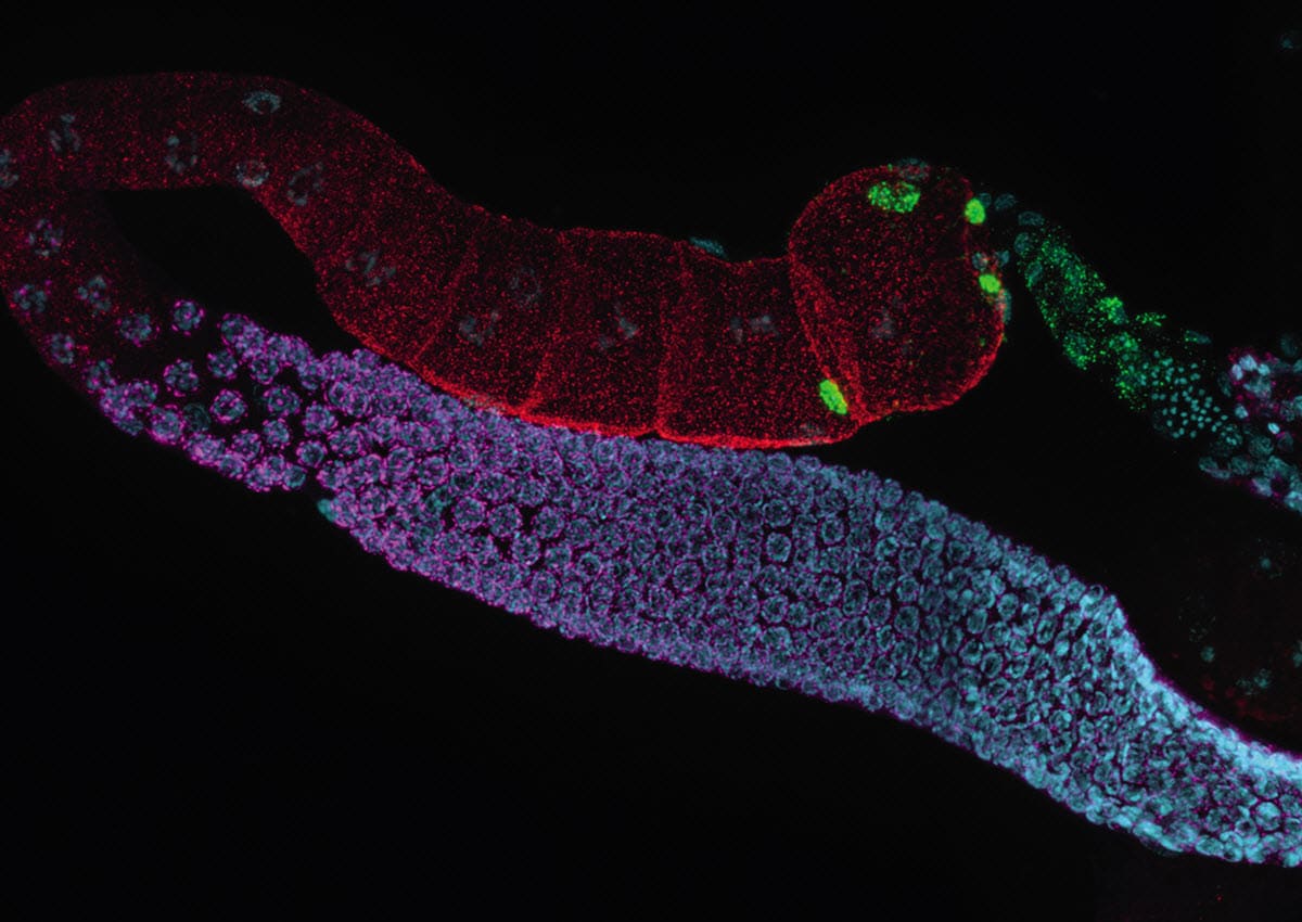
- THUNDER: Leica’s THUNDER Imagers offer Instant Computational Clearing (ICC) to generate high resolution, high contrast images, and reveal details which previously could not be observed.
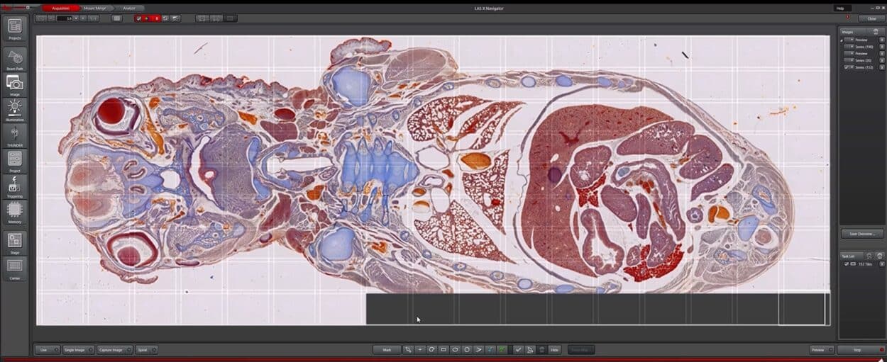
- Navigator: Explore your entire specimen with advanced specimen overview, scanning and stitching tools in Leica’s LAS X Navigator Software.