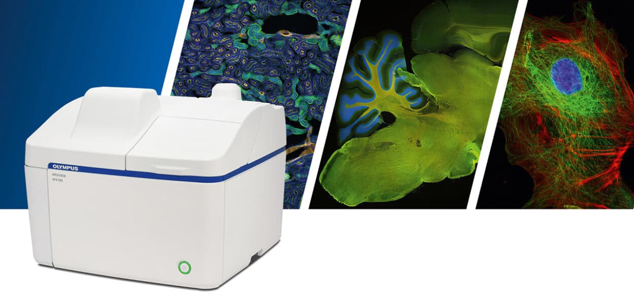

Capturing high quality microscope images is an integral part of life science but can be a time-consuming process once adjustments, focusing and image acquisition are taken into account. The APEXVIEW APX100 all-in-one microscope from Evident has been designed to streamline the microscope imaging workflow with automated image acquisition. Featuring renowned X Line optics, bespoke software, powerful AI and a suite of smart features, the APX100 system combines the usability of an all-in-one microscope with publication-quality image capture.
By Evident Europe.
Diverse Imaging Applications
The highly versatile APX100 microscope has been designed to support numerous imaging applications, with a wide range of holders that accommodate slides, dishes, well-plates and flasks.
The system can perform a wide range of imaging functions, such as multichannel, stitching, time-lapse, and Z-stack acquisition, as well as a variety of lighting options including brightfield, phase-contrast, fluorescence, and gradient contrast.
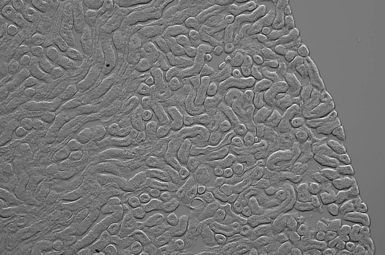
Developed by Evident, gradient contrast is a unique transmission observation method that enables the acquisition of sharp, high-contrast images (Fig 1).
Featuring the same X Line optics found in Evident’s high-end microscopes, it’s easy to achieve publication-quality imaging with the APX100.
Automated Features for Rapid Image Acquisition
Capturing high-quality images using a conventional microscope relies upon continuous fine-tuned adjustments to ensure that the subject is in-focus and the camera is optimally positioned. These time-consuming adjustments can account for a significant proportion of the imaging workflow, but with the APX100, these steps are automated.
Upon placing your sample in the sample holder, the APX100’s smart navigator takes a macro image, from which the AI software locates the sample and adjusts the stage to bring it into the center of the monitor display. From here, you can choose the observation method and immediately start capturing images. From loading your sample to image acquisition takes roughly 10 seconds.
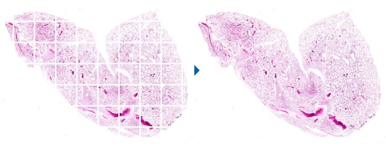
The APX100 makes imaging large samples easy thanks to automated scanning, high-speed autofocus and stitching features that enable you to capture a detailed image of your entire sample. APX100 achieves this by taking a series of high magnification images of each section of the sample, with autofocus adjusting between images.
The stitching function then combines these images into a single panoramic image displaying the whole subject (Fig 2).
When combined with the APX100’s Z-stacking function, image stitching can be used to capture high-quality images of thick samples in 3D.
A Compact, User-Friendly All-In-One Microscope
Exhibiting a small footprint, built-in anti-vibration technology and shielded optics, the APX100 can be placed anywhere. Since the system is a closed unit, imaging can be performed in a brightly-lit room with no effect on imaging, allowing imaging to be performed in a busy lab space, or in parallel with other experiments. Negating the need for a dedicated darkroom can save valuable space in your lab or core facility.
The platform is highly user-friendly, and requires minimal training—making the APX100 ideally suited for academic labs, as well as core imaging facilities. With the APX100, spherical aberration correction is automated with motorized correction collar adjustments—helping you to achieve sharp images every time.
Data management is simplified too—since the APX100 system automatically organizes and stores your data. When an image is taken, the software creates folders for each sample and saves the data into relevant folder. Furthermore, all image acquisition settings are saved with the images so that they can easily be recalled for future experiments.
Accelerate Your Image Acquisition With APX100
Featuring a suite of automated features and best-in-class optics, the APX100 makes capturing publication-quality images rapid and easy.
Want to accelerate your imaging workflow? Discover the value that APX100 could bring to your research.
Author: Laura Lleras Forero, Product Marketing Manager Life Science.