High-grade gliomas, including glioblastoma multiforme, are aggressive and malignant brain tumors that pose significant challenges in both diagnosis and treatment. The gold standard for high-grade glioma treatment1 is the usage of multi-modal therapy, consisting of surgery, chemotherapy and radiotherapy2.
Sponsored by Leica Microsystems.
The combination of these therapies aims to postpone tumor progression, extend patient survival and delay recurrence and patient death. The concomitant use of chemotherapy and radiotherapy can extend patient survival compared to radiotherapy alone3.
In more recent years, fluorescence-guided surgery has emerged as a promising and effective method to aid neurosurgeons in the visualization and resection of malignant tumors4.
Extending Tumor Resection Intra-Operatively
Intraoperative strategies have been developed to safely achieve this balance, improving patient outcomes and quality of life5. Neurosurgeons have several tools at their disposal such as surgical microscopes and intraoperative imaging tools: neuro-navigation, intraoperative MRI and intraoperative ultrasonography.
When it comes to neuro-navigation and diffusion tensor imaging (DTI), the risk of brain shift can be consequential and may lead to insufficient resection and neurological deficits6. Intraoperative MRI can help overcome that brain shift; however, it may lead to image distortion and inaccurate target registration and other limitations7.
Lastly, intraoperative ultrasonography can also help avoid brain shift, but the levels of anatomical details are much lower and residual disease can be difficult to detect8.
Fluorescence-guided surgery is a valuable tool that utilizes fluorophores and specific light wavelengths to better distinguish the metabolic patterns of the tissue for enhanced tumor resection. 5-aminolevulinic acid (commonly known as 5-ALA) is a component that has been introduced in the treatment of malignant gliomas9. 5-ALA must be administered to the patient orally 3 to 4 hours before surgery as the fluorescence peak occurs 6 to 8 hours after administration10.
The Benefits of 5-ALA Fluorescence-Guided Surgery
When performing surgery, the primary goal is to maximize the extent of resection while preserving as much functional brain tissue as possible.
Fluorescence-guided surgery can provide real-time guidance, allowing surgeons to make informed decisions about the extent of resection while the procedure is ongoing. When exposed to a malignant tumor, 5-ALA is converted into a fluorescent light, enhancing tumor visualization that helps the surgeon to better between unhealthy and normal brain tissue for better removal11.
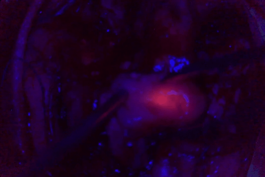
By aiding in the visualization of tumor margins, 5-ALA fluorescence allows for more complete removal of cancerous tissue during surgery. This is crucial for improving patient outcomes12.
A recent systematic review13 “found an observed increase in the overall survival (OS) and progression-free survival (PFS) of the 5-ALA group compared to the white light group, as well as an observed increase in the OS and PFS of complete resections compared to incomplete resections.”
Leica Microsystems is a pioneer in the field of fluorescence microscopy technology, developing innovative fluorescent modules for various medical applications.
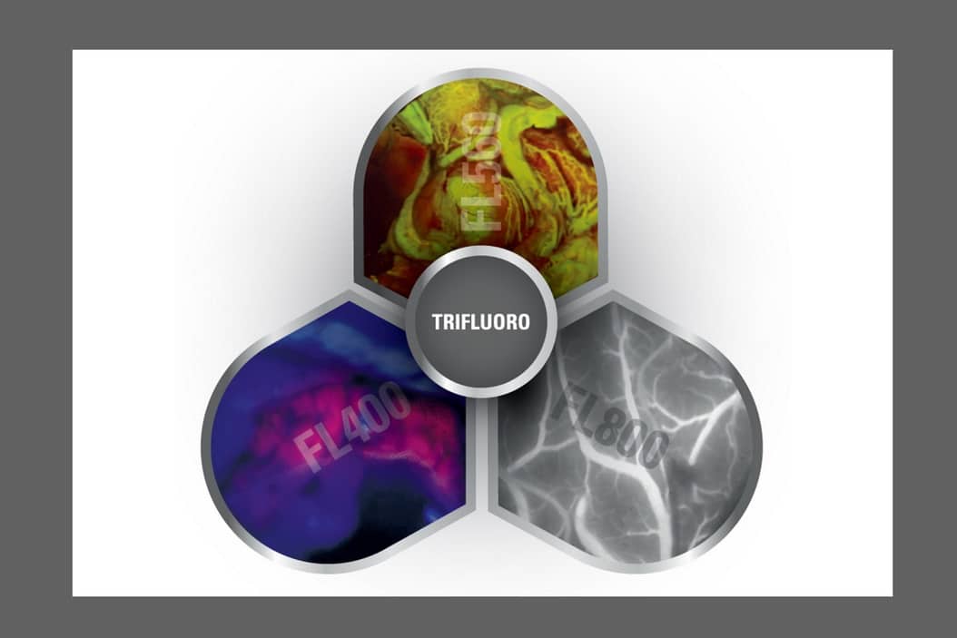
The ARveo 8 and M530 OHX neurosurgical microscopes from Leica Microsystems provide TriFluoro technology:
- FL400 fluorescence filter*: When used with 5-ALA, this filter supports resection procedures by allowing differentiation of tumor tissue from healthy brain tissue.
- FL560 fluorescence filter: Get a single, real-time view of fluorescent and non-fluorescent areas with clear differentiation and high contrast.
- FL800 intraoperative video-angiography module: When used in conjunction with ICG fluorescent dye, this feature allows neurosurgeons to clearly visualize blood flow in real-time.
*Please check with your local Leica Microsystems representative for product registration status.
During the Leica Microsystems Neurovisualization summit, Dr. Carla Reizinho, Neurosurgeon, Hospital Egas Moniz, Hospital da Luz of Lisbon, and Hospital da Luz of Oeiras, Portugal presented several clinical cases using 5-ALA for tumor resection. Watch the webinar on-demand here.
Discover our full range of Leica neurosurgical microscopes on MedicalExpo:
- Rong, L., Li, N. & Zhang, Z. Emerging therapies for glioblastoma: current state and future directions. J Exp Clin Cancer Res 41, 142 (2022). https://doi.org/10.1186/s13046-022-02349-7 ↩︎
- Dr Reizinho, C (2021) Intraoperative strategies to safely maximize extent of resection in High-grade Gliomas [Webinar]. https://www.leica-microsystems.com/science-lab/medical/surgical-management-of-high-grade-gliomas/ ↩︎
- Dr Reizinho, C (2021) Intraoperative strategies to safely maximize extent of resection in High-grade Gliomas [Webinar]. https://www.leica-microsystems.com/science-lab/medical/surgical-management-of-high-grade-gliomas/ ↩︎
- Mieog, J.S.D., Achterberg, F.B., Zlitni, A. et al. Fundamentals and developments in fluorescence-guided cancer surgery. Nat Rev Clin Oncol 19, 9–22 (2022). https://doi.org/10.1038/s41571-021-00548-3 ↩︎
- Haddad AF, Aghi MK, Butowski N. Novel intraoperative strategies for enhancing tumor control: Future directions. Neuro Oncol. 2022 Nov 2;24(Suppl 6):S25-S32. doi: 10.1093/neuonc/noac090. PMID: 36322096; PMCID: PMC9629473. ↩︎
- Dr Reizinho, C (2021) Intraoperative strategies to safely maximize extent of resection in High-grade Gliomas [Webinar]. https://www.leica-microsystems.com/science-lab/medical/surgical-management-of-high-grade-gliomas/ ↩︎
- Dr Reizinho, C (2021) Intraoperative strategies to safely maximize extent of resection in High-grade Gliomas [Webinar]. https://www.leica-microsystems.com/science-lab/medical/surgical-management-of-high-grade-gliomas/ ↩︎
- Dr Reizinho, C (2021) Intraoperative strategies to safely maximize extent of resection in High-grade Gliomas [Webinar]. https://www.leica-microsystems.com/science-lab/medical/surgical-management-of-high-grade-gliomas/ ↩︎
- Hadjipanayis CG, Widhalm G, Stummer W. What is the Surgical Benefit of Utilizing 5-Aminolevulinic Acid for Fluorescence-Guided Surgery of Malignant Gliomas? Neurosurgery. 2015 Nov;77(5):663-73. doi: 10.1227/NEU.0000000000000929. PMID: 26308630; PMCID: PMC4615466. ↩︎
- McCracken DJ, Schupper AJ, Lakomkin N, Malcolm J, Painton Bray D, Hadjipanayis CG. Turning on the light for brain tumor surgery: A 5-aminolevulinic acid story. Neuro Oncol. 2022 Nov 2;24(Suppl 6):S52-S61. doi: 10.1093/neuonc/noac191. PMID: 36322101; PMCID: PMC9629477. ↩︎
- Hadjipanayis CG, Widhalm G, Stummer W. What is the Surgical Benefit of Utilizing 5-Aminolevulinic Acid for Fluorescence-Guided Surgery of Malignant Gliomas? Neurosurgery. 2015 Nov;77(5):663-73. doi: 10.1227/NEU.0000000000000929. PMID: 26308630; PMCID: PMC4615466. ↩︎
- Front. Neurol., 16 June 2021 Sec. Neuro-Oncology and Neurosurgical Oncology Volume 12 – 2021 | https://doi.org/10.3389/fneur.2021.682151 ↩︎
- Eatz TA, Eichberg DG, Lu VM, Di L, Komotar RJ, Ivan ME. Intraoperative 5-ALA fluorescence-guided resection of high-grade glioma leads to greater extent of resection with better outcomes: a systematic review. J Neurooncol. 2022 Jan;156(2):233-256. doi: 10.1007/s11060-021-03901-9. Epub 2022 Jan 6. PMID: 34989964. ↩︎