![[MEDICA] Three Promising Trends in Medical Imaging](/wp-content/uploads/sites/9/MEDICA_Radiology_Trends.jpg)

Much of the technology on show at this year’s MEDICA 2022 relates to the field of radiology and medical imaging. MedicalExpo e-magazine caught up with Takashi Azuma, CTO of Japanese ultrasound imaging company Lily MedTech, and Sophia Borowka, CEO of Swiss ultrasound device company aiSon Technologies, to discuss some of the latest radiological trends and technologies that will be on display at MEDICA 2022. Both companies are participating in the event.
1/ Ultrasound Computed Tomography
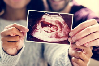
Many people are familiar with common medical imaging techniques such as x-ray tomography or magnetic resonance imaging (MRI). Most people are also familiar with b-mode ultrasound (gray scale imaging), which is typically used to produce images of growing fetuses.
A promising alternative is ultrasound computed tomography (USCT), which uses high-frequency sound waves emitted along a 2D slice of the human body to image the interior organs and bones. USCT is cheaper than MRI and there is no ionizing radiation involved, as there is with x-ray tomography.
Takashi Azuma said:
“Ultrasound imaging has developed greatly in terms of portability and ease of use in recent years. Attempts to use it in new fields, such as orthopedics, are now expanding beyond conventional diagnostic targets.
Difficulties controlling the accuracy of ultrasound imaging have been addressed with the evolution of USCT, which enables non-contact 3D imaging and records the entire necessary range. At present, breast cancer is still the main target, but the imaging of joints and new areas such as the brain are also being explored.”
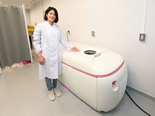
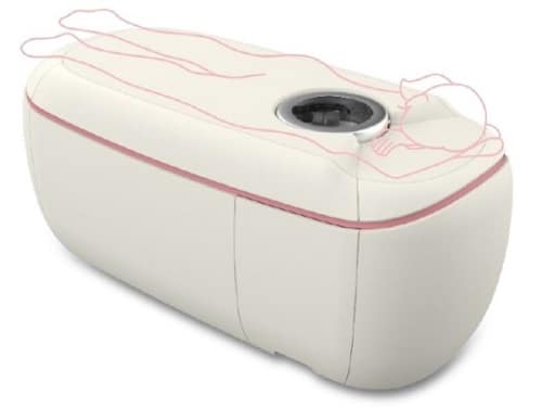
In 2021, Lily Medtech launched COCOLY, an innovative bed-type ultrasound examination device that uses a ring-array ultrasound transducer to scan for breast cancer.
2/ Handheld Ultrasound
Another important trend in ultrasound imaging is the move towards handheld devices, driven by advances in technology and the transition from inpatient to outpatient care. Today, the handheld ultrasound market is growing rapidly, as the latest generation of ultra-portable devices gains acceptance with medical professionals.
Sophia Borowka said:
“Thanks to intense research and technological advances, the image quality delivered by such devices is continually improving. Even superficial soft tissue structures, where ultrasound can trump CT and MRI imaging in terms of resolution and predictivity, can be readily imaged with specialized handheld ultrasound devices.”
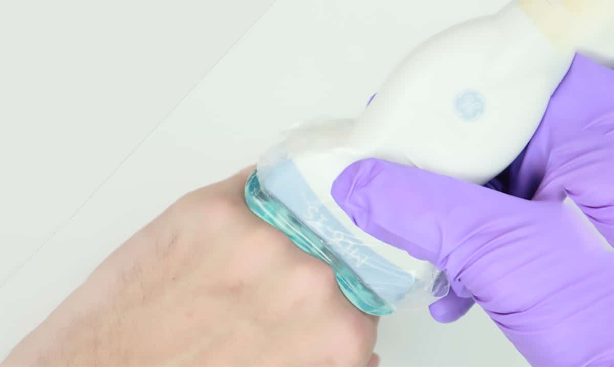
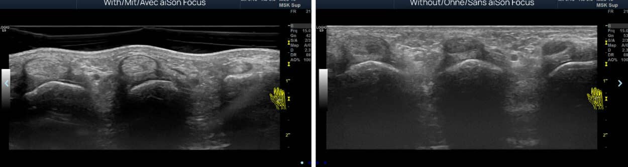
As handheld ultrasound technology has evolved, so have stand-off pads. By increasing the distance between the ultrasound transducer and the patient’s skin, such pads help to image superficial lesions, lesions located immediately below the skin, superficial foreign bodies, and breast lesions. She added:
“aiSon Technologies has now developed the first adaptive ultrasound pad. These reduce the amount of pressure that needs to be exerted on the patient’s skin, enabling an unobstructed view of the topmost anatomical structures. This can prevent wrong diagnoses in rheumatology and makes ultrasound-guided hand surgery much easier.”
3/ Next-Generation CADe and CADx
Traditional computer-aided detection (CAD) in mammography is nothing new—computers have been used to enhance the detection of mammographic abnormalities since the 1960s. Yet CAD has taken on a broader meaning more recently, notably in the context of renewed interest in artificial intelligence (AI).
Over the last decade, significant research has been conducted on deep learning. This has been made possible by advanced algorithms that are powered by faster computers, greater storage capabilities and the availability of big data.
New deep learning-based mammographic platforms, which differ from traditional CAD in several important ways, are expected to enhance radiological accuracy significantly. There are two components to consider: computer-aided detection (CADe), which focuses on detection, and computer-aided diagnosis (CADx), which focuses on classification. Compared with traditional CAD, next-generation systems will provide both CADe and CADx, offering a fully automated end-to-end process and better facilitating machine learning.
Takashi Azuma said:
“The development of AI, which eliminates the gap between the capabilities of non-specialists and specialists, has influenced many imaging modalities and extended the number of target diseases.
Going forwards, AI has the potential to not only learn from patient images with lesions, but also images from previous examinations, enabling the discovery of lesions that even skilled specialists cannot find.
Combined with the development of image-guided liquid biopsies and minimally invasive treatments, AI promises to revolutionize cancer detection, diagnosis and removal.”