
If you are looking for a surgical microscope for ENT surgery, you may be wondering how to make the best decision and maximize the value of your investment. In this article, we share some advice and considerations to help you make the right choice and ensure the equipment you select for the years to come meets your needs.
Content provided by Leica Microsystems
Some of the most common otolaryngology procedures require the use of an ENT surgical microscope, including neurinoma surgery, otosclerosis surgery, schwannoma surgery, cholesteatoma surgery, cochlear implant surgery, stapedectomy, tympanoplasty, laryngoplasty, and myringoplasty.
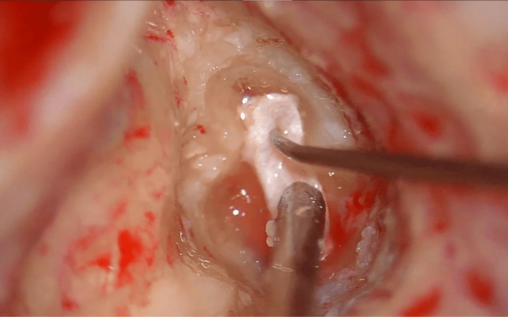
One of the major challenges is maintaining sufficient illumination while navigating complex anatomical structures and deep, narrow cavities. Proper illumination during ENT surgery is crucial for accurate location and surgical site access while minimizing iatrogenic injury to nearby structures and tissues.
In addition, during ENT surgical procedures, medical professionals typically work in different operating positions and can be positioned closely to the operating field. As such, they require a microscope with strong adaptability and intuitive operation.
Here are some key factors to consider when purchasing an ENT microscope:
1/ Illumination
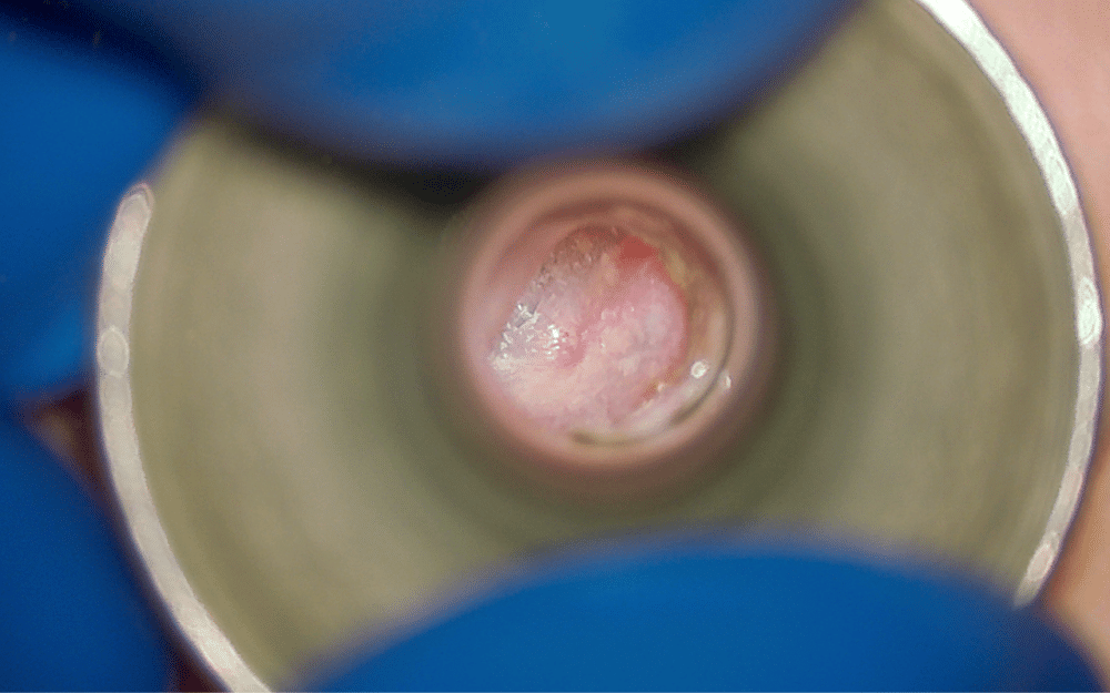
Illumination is critical to work in deep and narrow cavities. Microscopes with premium apochromatic optics and tailored illumination provide a crisp, true-color image even at the bottom of the deep, narrow channels typical of ENT surgery. They can also allow to visualize deep cavities during non-surgical procedures such as placement of transtympanic aerator or foreign body removal. Features such as automatic light intensity adaptation support patient safety, avoiding damage to sensitive tissues.
2/ Magnification
High magnification allows ENT specialists to easily access and target small, delicate structures. This is critical in stapes surgery for example, as the structure in the middle ear around the stapes measures less than two millimeters. ENT microscopes should provide strongly magnified high-quality images.
University of Bordeaux in France
3/ Depth of Field
Good depth of field provides a clear, three-dimensional view of the surgical field. This is particularly important when performing surgery in narrow spaces, for example in trans-temporal routes going to the CP angle or the internal canal.
4/ Adaptability
An adaptable microscope supports seamless operation during procedures and surgeon comfort. Features augmenting adaptability include positioning freedom and easy maneuverability allowing smooth movement of the microscope with one hand; adaptable binoculars that can be positioned and angled in order to fit specific operating requirements; intuitive and easy-to-reach microscope controls, including programmable footswitch and handles as well as resistance to vibrations and sturdy design.
5/ Integration
The integration of ENT surgical microscopes with other devices can allow to meet a wide spectrum of requirements in clinical use and teaching. Leica Microsystems for example offers an assortment of product accessories for increased efficiency and smooth workflows. This includes binoculars and objective lenses, single or dual handles, imaging and recording systems, stand-mounted monitors, side observers, micromanipulator for CO2 laser surgery.
Selecting a microscope for ENT surgery requires some thought to ensure you make the right choice for your clinical setting. Exchanges with manufacturers can help you better understand the differences between the microscopes on the market, and which one best fits your requirements. A product demonstration is also a good idea to see the microscope in operation. This will help you confirm your decision, ensuring the microscope you have chosen meets your objectives.
Interested in ENT Surgery and Consultation Microscopes By Leica Microsystems
Leica Microsystems offers a wide range of solutions for ENT specialists to support both ENT surgery and consultations. Our innovative technologies provide enhanced depth of field, precise positioning, and great maneuverability and mobility. Discover the range here.
Uniquely from Leica Microsystems
BrightCare Plus* technology automatically adapts light intensity according to the microscopes’ working distance to prevent damage to sensitive tissues.
The BrightCare Plus feature allows maximum illumination at long working distances. As working distance decreases, the light intensity is reduced automatically, diminishing the risk of tissue damage and burns (up to 60% reduction of light intensity). Conversely, the light intensity increases proportionally with working distance.
BrightCare Plus is also available on both the M530 OHX and PROvido microscopes.
*BrightCare Plus is available in the M530 OHX Microscope and PROvido microscope for ENT surgery