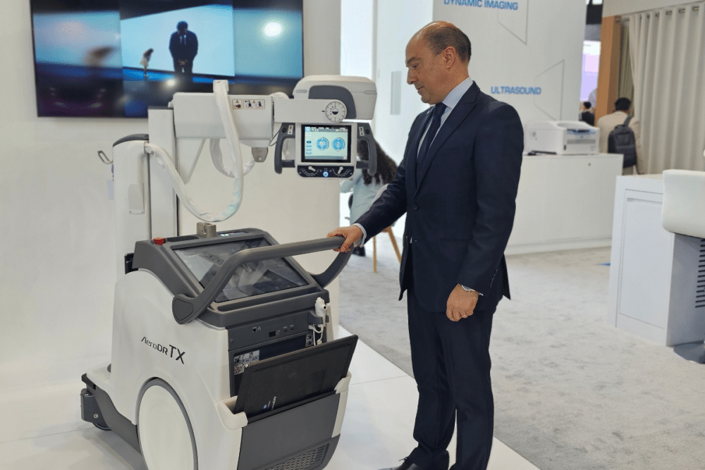

At Arab Health in Dubai last week, we spoke with Mário Garcia, Marketing Manager EMEA & Regional Manager EW at Konica Minolta, about their new mobile radiography device, the AeroDR TX. This product is revolutionizing radiography by bringing radiology to the patient’s bedside with dynamic images and AI, therefore transforming diagnostics. It won second place in the i-NOVO Award for medical innovation.
Can you present the AeroDR TX?
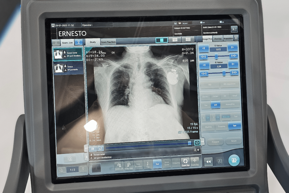
Mário Garcia: “The AeroDR TX is the only mobile radiography device capable of capturing dynamic images. With this system, we can bring dynamic images directly to the patient’s bedside. It enables the acquisition of dynamic images at 15 frames per second, allowing real-time visualization of X-ray exams—something that was previously impossible with static images.”
Why is it mobile?
Mário Garcia: “The AeroDR TX is a mobile that brings diagnostics directly to the patient location allowing the doctors to have a new way of making X-ray diagnostics. In some procedures, there is no longer a need to transfer the patient to a CT scanner or even to the radiology department. For example, in an ICU, patients are often intubated and in critical condition, making movement difficult. With this solution, the device can be brought to the patient’s location, whether in nursing units, ICUs, or rehabilitation departments.”
How does the imaging process work?
Mário Garcia: “This system includes a dedicated dynamic wireless flat panel that captures X-ray images. The console of the AeroDR TX allows the control of imaging parameters, such as kilovoltage (kV) and milliampere-seconds (mAs), which determine radiation dosage. These images are then processed on the CS-7 software workstation of the mobile unit, significantly improving diagnostic accuracy.”
Who operates the device? Nurses, doctors, or radiologists?
Mário Garcia: “The radiographer operates the device and brings it to the patient’s location.”
Which body parts can it be used for?
Mário Garcia: “It can be used for all body parts, but the dynamic imaging capability is particularly useful for lung imaging and orthopedics. It allows visualization of movements, such as the motion lungs, arm or knee, making it valuable for multiple applications.”
Since X-rays involve radiation, does this require a specific shielded area?
Mário Garcia: “In a standard X-ray room, dedicated shielding (such as lead walls) is required. However, the AeroDR TX operates with a much lower radiation dose compared to a conventional X-ray room, so such shielding is not necessary. Compared to a fluoroscopy exam, where the patient is exposed to continuous radiation, the AeroDR TX reduces radiation exposure by at least 30% depending on the exam type.”
Despite the lower dose, does it still provide high-quality images?
Mário Garcia: “Yes, thanks to advanced algorithms and panel technology. The system captures image slices during the X-ray procedure, reducing the required radiation dose compared to a CT scan, which typically requires multiple high-dose exposures.”
What about the duration of the exam?
Mário Garcia: “The exam is significantly faster. This also results in cost savings, as patients no longer need to be transported to a CT scanner or fluoroscopy system. Instead, a single person can operate the mobile unit and perform the exam near the patient.”
What kind of preparation does the patient need?
Mário Garcia: “No special preparation is required. The process is identical to a standard X-ray, but with the added benefit of dynamic imaging.”
Do users require special training?
Mário Garcia: “Yes, radiographers need to be trained on how to acquire images with the system. Additionally, if artificial intelligence (AI) is used, radiologists must learn how to interpret AI-assisted images in our advanced AI software.”
How does this benefit radiologists? What new insights does it provide?
Mário Garcia: “Dynamic imaging provides crucial insights that static imaging cannot. For instance, it helps determine whether a pacemaker catheter is correctly positioned, ensuring its proper placement. Additionally, it plays a key role in assessing lung function by evaluating the patient’s breathing capacity. While a static image may suggest that everything appears normal, dynamic imaging reveals whether the lungs are functioning correctly and if the patient is breathing properly. This system therefore enables radiologists to detect new diagnostic insights that were not previously possible with static imaging. New software tools with AI are being developed and incorporated in our Dynamic Radiography technology.”
What happens after the images are acquired?
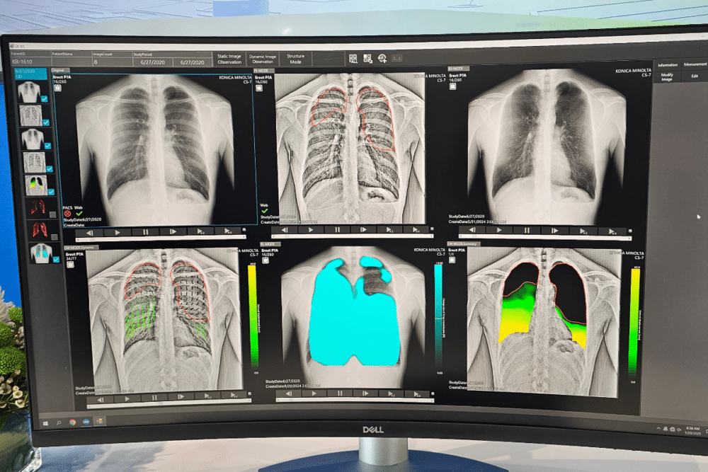
Mário Garcia: “The dynamic images are analyzed using artificial intelligence. From these images, different diagnostic views can be generated, helping assess lung function, areas of reduced capacity, and abnormalities. For example, in one acquisition, green areas may indicate missing lung capacity. Another image can provide lung measurements and breathing assessments, etc.”
Can radiologists customize the AI parameters?
Mário Garcia: “Yes, AI provides an initial diagnosis, but the radiologist has full control to modify parameters. If they disagree with the AI’s assessment, they can adjust the algorithm, which then learns and improves for future cases—this is the core of deep learning technology.”
Do radiologists need training in AI?
Mário Garcia: “AI-assisted diagnostics require a new way of interpreting medical images. Radiologists are still adapting to this technology and learning how to integrate AI into their workflows. Over time, the software will improve based on their feedback, continuously refining its accuracy.”
Is this technology recognized in the medical community?
Mário Garcia: “Yes, it has been extensively studied and published. Several clinical cases have been presented at the Radiological Society of North America (RSNA) and ECR (European Congress of Radiology), and a significant body of research supports its effectiveness.”
How many units does a hospital typically need?
Mário Garcia: “This is a new and rapidly growing technology. We have already established key opinion leaders in Italy and Spain, with additional units installed in Belgium, Germany, Qatar, Saudi and Egypt. The technology is expanding quickly, as there are currently no direct competitors.”