
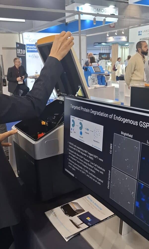
Promega’s GloMax® Galaxy Bioluminescence Imager enables real-time visualization of protein dynamics and cellular processes using NanoLuc® luciferase technologies.
We have rewritten the original article published on March 26 to ensure the accuracy of technical details. Here are the main highlights to know about the GloMax Galaxy Bioluminescence Imager by Promega:
- New bioluminescence microscope enables functional imaging of NanoLuc® technologies in living and fixed cells.
- The GloMax® Galaxy also offers fluorescence and brightfield imaging capabilities.
Promega’s GloMax® Galaxy Bioluminescence Imager, showcased at Forum LABO Paris 2025, is revolutionizing cellular imaging by providing researchers with real-time, high-sensitivity bioluminescence visualization. Unlike traditional imaging systems that rely on external excitation, the Galaxy Imager is specifically designed to detect self-emitted light from luciferase-based reactions, enabling low-background, high-contrast imaging of live cells with minimal phototoxicity.
“Bioluminescence is an enzymatic reaction in which the enzyme luciferase, in the presence of its substrate, produces light,” a representative for Promega explained during the event. “This phenomenon occurs naturally in certain animals, such as fireflies and deep-sea marine creatures like shrimp and jellyfish, as well as in some bacteria.”
Inspired by this natural process, Promega has developed numerous assays and cellular tests based on this technology.
Advantages of Bioluminescence Imaging
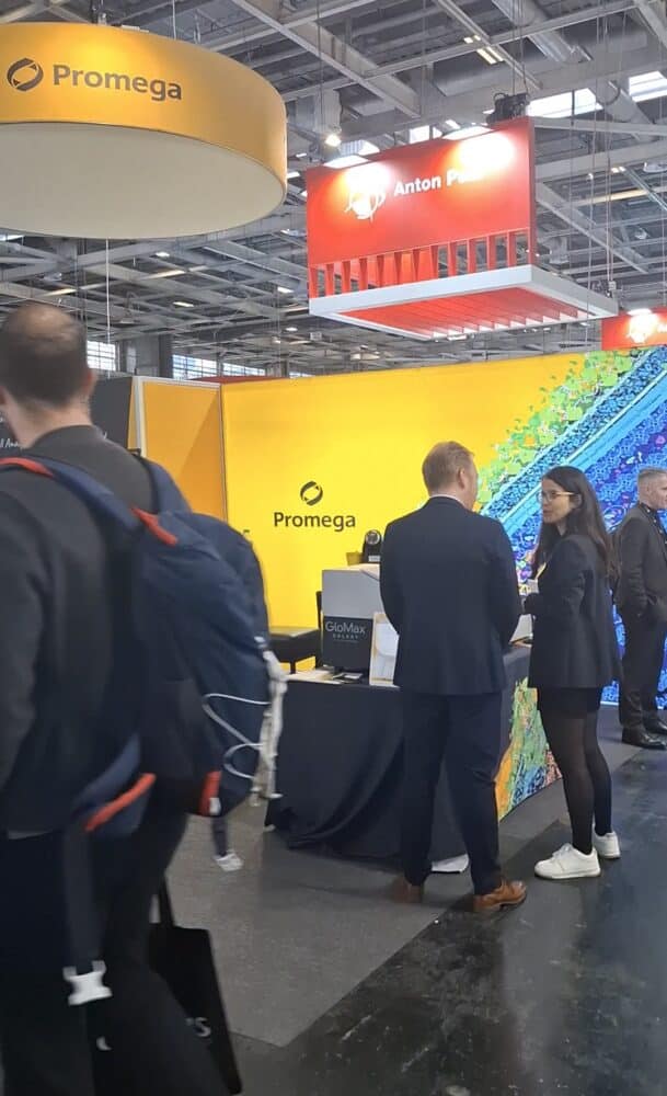
At Forum LABO Paris 2025, Promega introduced the GloMax® Galaxy for the first time at a trade show in France.
“Initially, Promega offered plate readers to quantify bioluminescence signals in Relative Light Units (RLU). However, these devices could only measure signal intensity and not visualize what was happening inside the cells.”
Bioluminescence imaging provides several key advantages over fluorescence imaging. Its naturally low background noise allows for the detection of weak photon signals and low-abundance proteins with high sensitivity.
By generating a stable and sustained signal on its own through bioluminescence, the system reduces the risks of phototoxicity and photobleaching. This makes it possible to conduct repeated imaging sessions over long periods without harming cells or altering their physiological state. The Galaxy system enhances research by offering spatially resolved imaging of protein localization, intracellular trafficking, and protein-protein interactions, complementing quantitative bioluminescence data obtained from plate readers. While primarily optimized for NanoLuc® luciferase-based assays, the system also supports multiplexing with fluorescence imaging, allowing researchers to overlay fluorescent markers for more comprehensive cellular analysis.
Observing Biological Processes at a Single-cell Level
The GloMax Galaxy Bioluminescence Imager bridges the gap between luminescence quantification and visualizing cellular events at the single-cell level. With this innovation, researchers can now move beyond bulk signal measurements to directly observe biological processes, opening new possibilities for studies in live-cell imaging, drug discovery, and protein dynamics. By integrating cutting-edge bioluminescence technology, Promega continues to push the boundaries of cellular biology research.
“While primarily designed for bioluminescence imaging, the Galaxy system also integrates fluorescence imaging, enabling bioluminescence-fluorescence multiplexing. For instance, in an image, researchers can visualize an intracellular bioluminescent signal and use fluorescence to stain the nucleus. By overlaying both signals, they can analyze cellular structures in greater detail.”
As for the relationship between Promega’s imaging systems, the Galaxy and plate readers are separate devices but work in a complementary manner. Researchers typically first quantify bioluminescence signals using a plate reader—whether Promega’s or another luminometer—and then use the Galaxy Imager for spatial visualization at the cellular level. This distinction is crucial: a plate reader may indicate a strong bioluminescence signal, but without imaging, it is impossible to determine whether all cells are producing the signal or if it originates from a small subset. The Galaxy Imager helps researchers differentiate whether 10%, 20%, or more of the cells are responding, which can be critical in applications such as drug development and therapeutic research. If only a small percentage of cells respond to a treatment, scientists may need to adjust their approach accordingly.