Funded by the Carl Zeiss Foundation, the OptoCarDi project is developing a multimodal high-resolution imaging catheter that will revolutionize myocarditis diagnosis. This unique optical system will be able to identify the chemical composition, molecular information and structural morphological changes in the myocardium in vivo, instantly recording all parameters on the spot. Significant breakthroughs are also expected in the treatment of heart disease in general.
Inflammation of the heart muscle—the myocardium—is known as myocarditis. This condition can be caused by a number of different factors, such as viruses, drug reactions or general inflammatory conditions.
A heart affected by myocarditis has reduced pumping capacity and patients experience symptoms such as chest pain, shortness of breath and rapid or irregular heart rhythm (arrhythmias). Heart inflammation can also result in clots, which can lead to strokes or heart attacks.
The treatment for myocarditis includes medication, minor procedures and even major surgery. Prior to that, the condition and its cause need to be properly diagnosed. When a patient’s recent medical history and the results of exams such as an echocardiogram or magnetic resonance imaging (MRI) are not conclusive in determining the most likely cause of the myocarditis, the gold standard diagnostic tool is a biopsy.
However, this can be a problematic solution: while a single procedure of that kind has low sensitivity, performing multiple procedures to collect numerous samples of tissue is associated with many complications. That is where OptoCarDi comes in.
Collosal Technical Challenges
The aim of the project is to develop a multimodal high-resolution imaging catheter that will be able to identify the chemical composition, molecular information and structural morphological changes in the myocardium in vivo (while the heart chambers are fully perfused), instantly recording all parameters on the spot.
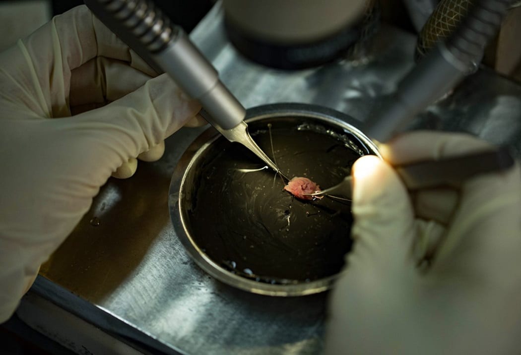
To make this achievable, the catheter probe will be the first to integrate three different sensor technologies: optical coherence tomography, UV-excited autofluorescence and near-infrared spectroscopy. The device is to capture the structure of the heart based on optical signals—like a microscope, but inside the body.
The technical challenges involved are considerable. Iwan Schie, Professor of Biomedical Engineering at Ernst-Abbe-Hochschule Jena at EAH (University of Applied Sciences Jena), told MedicalExpo e-magazine:
“It’s crucial for the probe’s outer diameter to remain small, with only a few millimeters, as this will ensure its compatibility with intracardiac insertions. Moreover, maneuverability is a key consideration; the probe should be easily navigable within the heart by the operator.
These requirements come with their own set of challenges. Crafting specific optical systems, which includes the creation of specialized lenses and optical fibers, is a pivotal component of our undertaking.
Additionally, we’re tasked with developing a unit that not only facilitates the coupling of the excitation light from the modalities but also effectively handles the corresponding detection signals. Achieving this without incurring significant losses or crosstalk is technically demanding.
Lastly, given the dynamic environment of the heart, our external data acquisition system must be adept at rapid data collection. This speed is essential to prevent motion artifacts during in vivo measurements of the heart. Complementing this, the software codes designed for processing the gathered data must prioritize computational efficiency and speed, ensuring real-time or near-real-time analysis.”
Treatment of Heart Disease in General
Patients with other heart conditions will also benefit from the OptoCarDi’s catheter. Significant breakthroughs are also expected in the treatment of heart disease in general as the device will allow for monitoring that does not involve tissue samples.
Prof. Iwan Schie explained:
“The system also excels in pinpointing pathologies such as those manifesting as fibrosis or scarring from prior conditions like heart attacks.
Additionally, it can detect myocardial necrosis arising from coronary heart disease, atherosclerosis or diabetes-induced hypoxia. Also, it can identify and characterize changes in heart tissue due to leukocyte infiltration without the use of markers and without causing damage.
Moreover, its unique approach to examining the inner recesses of the heart chambers allows for a detailed assessment of the endocardium. This inherent capability is invaluable, especially when there’s a need to detect problems like endocarditis, a condition which can be elusive in its early stages.”
As one of the oldest and largest private science-funding organizations of its kind in Germany, the Carl Zeiss Foundation is funding the project, which commenced in June 2023 and will continue until May 2026. The goal is to prepare the laboratory prototype for preclinical testing and to approve the technology for use on patients.
The research team is led by Prof. Schie and Prof. Dr. Robert Brunner (Miniaturized Optical Sensor Systems), with their groups from EAH—plus cardiologists Prof. Sven Möbius-Winkler and Prof. Christian Schulze, both from the Jena University Hospital (UKJ).
The work will require close collaboration of experts from different disciplines such as optics, biomedicine and medicine. Among industry partners to be included are Grintech GmbH in Jena (the global leading manufacturer of the gradient-index optics to be integrated into the sensor head), the Leibniz Institute of Photonic Technology (Leibniz-IPHT) Jena for the development of optical fibers and the Liryc Institute in Bordeaux, well-known in cardiology research.
.

